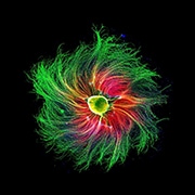
Each year since 1975, Nikon’s Small World has celebrated images captured by the light microscope. In their 2021 Photomicrography Competition, Nikon awarded fourth place to Paula Diaz, a PhD candidate in the Department of Physiology at Pontificia Universidad Católica de Chile in Santiago. Using fluorescence microscopy and 10x objective lens magnification, Diaz captured a colorful image of a rat embryo’s dorsal root ganglion. The image was also recognized by Nature as one of its best science images of 2021. In addition to Paula’s work, Nikon’s Small World also honored Andrea Tedeschi, PhD, The Ohio State University, for capturing the 3D vasculature of an adult mouse somatosensory cortex using confocal microscopy and 10x objective lens magnification. To view these incredible images, and see other amazing content, be sure to visit Nikon’s Small World or Nature.
Image credit: Paula Diaz, Pontificia Universidad Católica de Chile/Nikon Small World.


