The role of the amygdala in pain has been closely considered by pain researchers for decades. The results have been somewhat contradictory: Recent research has mostly found pronociceptive contributions of the amygdala, but earlier studies revealed an antinociceptive function. Now, a new study brings clarity to the question of how the amygdala, an almond-shaped structure deep in the temporal lobe of the brain, regulates pain.
Researchers led by Yarimar Carrasquillo, National Institutes of Health, Bethesda, US, find that the central nucleus of the amygdala (CeA) contains two distinct populations of neurons that bidirectionally modulate pain in mice. In particular, CeA neurons that express protein kinase C-delta (CeA-PKCδ) are pronociceptive, and become sensitized and drive pain-related behaviors in a mouse model of neuropathic pain. Meanwhile, CeA neurons that express somatostatin (CeA-Som) are antinociceptive and become inhibited during nerve injury-induced pain.
Pain researchers had previously suspected that bidirectional control of pain behaviors could be driven by different cell types in the amygdala, but until the current study, the mechanisms had remained unknown, said Ben Kolber, a pain researcher at Duquesne University, Pittsburgh, US, who was not involved in the new research.
“The cell type-specific data in these studies showing opposing whole animal effects of amygdala modulation are quite exciting,” Kolber said. “This study confirms a long-outstanding hypothesis in pain research and will help propel the field by ‘bringing back’ the importance of the analgesic side of the conversation,” he said.
The study was published October 8, 2019, in Cell Reports.
“A smaller scalpel”
“For most of my career,” said Carrasquillo, explaining the background to the new study, “I’ve been interested in understanding how the brain drives pathological pain. When we started this project, I wanted to know the maladaptive changes that happen at the neuronal level to drive pain,” according to Carrasquillo, who set her sights specifically on the amygdala.
The amygdala receives converging sensory inputs along with affective inputs from higher brain structures, integrating all of that information and converting it into behavioral outputs. It is this convergent processing that likely drives pain’s affective components. This has led to the view that the amygdala promotes pain, but the reality is more complicated.
“The focus of research on the amygdala’s role in pain in the past 15 years has been that the amygdala is the site of pronociception,” Carrasquillo told PRF. “I was puzzled, because I knew papers from the 1980s and 1990s that showed that the amygdala is also involved in analgesia. That’s what made me think that maybe pronociception and antinociception happen in a cell type-specific manner. Maybe when we manipulate all cells of the amygdala we only get to see one side of the story, but perhaps if we use a smaller scalpel we can tease out more bidirectional control of pain.”
In pursuit of that “smaller scalpel,” Carrasquillo and colleagues, including co-first authors Torri Wilson, Spring Valdivia, and Aleisha Khan, used genetic approaches in mice to fluorescently label and manipulate two independent populations of neurons in the CeA that have little overlap of genetic markers. These were the CeA-PKCδ neurons and the CeA-Som neurons.
The investigators first asked whether these two neuronal populations each received direct sensory input from the parabrachial nucleus, a brainstem nucleus that relays sensory input from the spinal cord to higher brain regions. They used optogenetic techniques to stimulate parabrachial nucleus nerve terminals in the CeA while recording CeA neuronal activity in acute brain slices. The results showed that, in most of the labeled CeA-PKCδ and CeA-Som neurons, light stimulation evoked excitatory postsynaptic currents (EPSCs), showing that both populations received excitatory input from the parabrachial nucleus.
The authors then tested what happened to the CeA-PKCδ and CeA-Som neurons during pain-related behaviors by using a sciatic nerve cuff model of neuropathic pain in mice. As expected, nerve-injured animals showed increased hypersensitivity to thermal, tactile, and pinch stimuli, compared to sham-operated animals. This was accompanied by substantial activation of CeA-PKCδ neurons but only minimal activation of CeA-Som neurons (as measured by levels of the activated form of extracellular signal regulated kinase, or ERK, as well as c-Fos expression―two surrogate markers of neuronal activity). Similarly, patch clamp recordings from acute brain slices revealed that CeA-PKCδ neurons were electrically hyperexcitable in nerve-injured mice, particularly late-firing neurons.
Together, the results showed that pain-related behaviors were accompanied by activation specifically of CeA-PKCδ neurons, suggesting that perhaps these cells were pronociceptive contributors in the neuropathic pain model.
The yin and yang of pain modulation in the amygdala
To test that hypothesis, and in particular to show a causal role in pain for the CeA-PKCδ neurons, Carrasquillo and colleagues used a chemogenetic technique that allowed them to selectively excite or inhibit CeA-PKCδ or CeA-Som neurons. Inhibition of CeA-PKCδ neurons in nerve-injured animals caused a dramatic reversal of tactile and thermal pain hypersensitivity, while activation of these neurons was sufficient to promote tactile, though not thermal, hypersensitivity in the absence of nerve injury. These results showed that CeA-PKCδ neurons were pronociceptive, with the behavioral modality they affected dependent on the context.
But what about the CeA-Som neurons? While the evidence had suggested that these neurons received sensory input via the parabrachial nucleus, few seemed to be activated following nerve injury. Patch clamp recordings from CeA-Som neurons in acute brain slices indeed revealed reduced spontaneous activity following nerve injury.
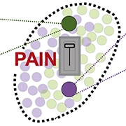
![Proposed Model for Dual and Opposing Modulation of Pain-Related Behaviors in the CeA. The CeA functions as a pain rheostat, attenuating or exacerbating pain-related behaviors in mice. The dual and opposing function of the CeA is encoded by opposing injury-induced changes in the excitability [of] CeA-PKCδ (green, left) and CeA-Som (purple, right) cells, with increases in firing in CeA-PKCδ neurons and attenuation of excitability in CeA-Som cells following injury. Activation of these two populations of CeA cells promotes completely opposite behaviors, with activation of CeA-PKCδ cells driving increases in pain (pronociception) and activation of CeA-Som neurons resulting in decreases in pain (antinociception). Image and caption from Wilson et al. Cell Rep. 2019 Oct 08; 29(2):332-346.e5. Creative Commons Attribution – NonCommercial – NoDerivs (CC BY-NC-ND 4.0).](https://www.iasp-pain.org/wp-content/uploads/2023/02/AmygdalaInline.jpg)
Carrasquillo then tested whether the CeA-Som neurons modulated pain behavior. Using chemogenetics to manipulate the activity of these neurons, the authors found that inhibition of CeA-Som neurons was sufficient to cause tactile hypersensitivity in the absence of nerve injury. Moreover, activation of CeA-Som neurons reversed tactile hypersensitivity from nerve injury.
“One of the really interesting bits of data is that the CeA-Som neurons are inhibited in the context of painful injury,” Kolber said. “These data fit with our findings from a few years ago that there is an ongoing antinociceptive signal from the CeA,” he said, referring to work published in 2017 (Sadler et al., 2017).
All in all, the experiments showed that the CeA exerts pronociceptive effects via CeA-PKCδ neurons, and antinociceptive effects via CeA-Som neurons. Nerve injury shifts the balance of CeA activity to favor pronociception by sensitizing CeA-PKCδ neurons and decreasing the activity of CeA-Som neurons.
Carrasquillo said she was extremely grateful to learn that the two different CeA neuronal populations had such clearly defined and separate roles in pain modulation, even though the cells are not anatomically segregated in the CeA.
“We have identified a structure in the brain where cells are intermingled and yet have totally opposite outputs. We show that these cells have a yin-yang type of relationship where there are increases in excitability in one population and decreases in excitability in another population,” she said.
When asked what future directions her lab will explore, Carrasquillo chuckled and said that she always tells people it is a waste of an amygdala to study only the somatosensory component of pain.
“One of the things we’re looking at is the inputs and outputs that are modulating the affective component of pain,” she said.
Kolber also stressed the importance of investigating that aspect of pain.
“It would also be interesting to know the impact of these cells on pure aversion. In other words, would driving CeA-PKCδ neurons cause context aversion in a real-time place preference/aversion behavior assay?” he said. "As knowledge of these intra-amygdala circuits is defined, we may ultimately understand issues related to psychiatric disturbances in chronic pain.”
Fred Schwaller, PhD, is a postdoctoral researcher at the Max Delbrück Center for Molecular Medicine in Berlin, Germany.


