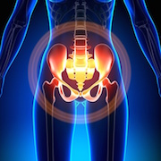
On June 6, 2017, PRF hosted its 26th webinar, this one devoted to the topic of chronic pelvic pain. Julie Christianson, University of Kansas, Kansas City, US, delivered a presentation based on her work and that of others investigating the nerves that innervate visceral organs, how they produce abdominal and pelvic pain, and how comorbidity with other pain conditions and mood-related disorders might arise. Stress—particularly early in life—emerged as a critical factor.
The webinar was moderated by Richard Traub, University of Maryland, Baltimore, US, who was joined by panelists Jennifer DeBerry, University of Alabama at Birmingham, US, and Benedict Kolber, Duquesne University, Pittsburgh, US.
To begin, Christianson said, “It’s important to note that chronic pelvic pain itself is a symptom, rather than a syndrome,” but it is associated with several so-called “functional pain disorders”—syndromes of unknown etiology that feature chronic and often widespread pain. These include irritable bowel syndrome (IBS), interstitial cystitis (IC; also called painful bladder syndrome), vulvodynia, and chronic prostatitis/chronic pelvic pain syndrome (CPP). Each of these affects 4 to 20 percent of the US population.
Patients with these syndromes (and functional pain disorders in general) display high comorbidity with other pain conditions and with mood disorders. Stress is a crucial aspect, as patients are often more susceptible to it, and stress can trigger painful episodes. Also, “Far more women are diagnosed with these syndromes than are men,” Christianson said, which may be due to hormonal differences.
Signals from within
Somatic innervation of the skin has been clearly described, with specific spinal segments sending afferents to clearly delineated parts of the dermatome. “It’s very easy to identify pain in structures on the outside of the body,” Christianson said. “Unfortunately, when we talk about structures inside the body, things get a bit more muddled.”
Although sensory neurons that innervate the visceral organs are found in the dorsal root ganglia (DRG) and nodose ganglia, the afferent sensory nerves travel alongside efferent nerves of the sympathetic and parasympathetic branches of the autonomic nervous system. Christianson, working at the time as a postdoc in the lab of Brian Davis at the University of Pittsburgh, US, used retrograde tracing to get a better picture of which nerves serve which abdominal organs. She found that, unlike with more restricted dermatomal distribution, the organs were all served by nerves originating from multiple spinal levels with a loose rostrocaudal afferent innervation organization maintained at their spinal inputs (e.g., Christianson et al., 2007). Ongoing work in Christianson’s own lab has since found a similar rostrocaudal organization of innervation of pelvic organs. The innervation by multiple spinal segments makes pain hard to localize, likely contributing to comorbidity.
Unlike nerve endings in the skin that may innervate specialized end-organs, research suggests that visceral nerve endings are “free,” but display various arborization patterns. Further complicating studies of visceral nerve endings are the abundant nerves of the separate enteric nervous system that resides within the gut.
Afferent nerves from visceral organs have been categorized based on their responsiveness to mechanical stimuli including probe and stretch, and on their firing patterns (Brierley et al., 2004). In a study from Davis’ group (Malin et al., 2009), about a third of afferent neurons from the colon responded to colon distension with high-frequency firing that tracked the stimulus strength, whereas the other two-thirds fired at low frequency in response to any stimulus.
The populations also differed in neurochemical phenotype: The low-frequency firing afferents, which are implicated in inflammatory visceral pain and the development of functional pain disorders, expressed transient receptor potential receptor vanilloid type 1 (TRPV1) and glial cell line-derived neurotrophic factor receptor alpha 3 (GFR-α3), whereas the high-frequency firing neurons did not. Earlier work by Christianson in the Davis lab (Christianson et al., 2006) showed that afferents innervating the colon were enriched in TRPV1 and calcitonin gene-related peptide (CGRP)—markers of peptidergic neurons—whereas there was little evidence of myelination or isolectin B4 (IB4) labeling.
Ben Feng in Gerald Gebhart’s lab, also at the University of Pittsburgh, then found a third population of “silent” neurons in the colon that responded to electrical but not mechanical stimulation. Some of these neurons became mechanically sensitive following exposure to inflammatory molecules (Feng and Gebhart, 2011).
A different study from Gebhart’s lab of mRNA in individual visceral afferents from the bladder showed that neurons at the lumbosacral region of the spine did not contain TRPA1, but neurons at other spinal levels contained TRPV1 and TRPA1 (La et al., 2011). And a study from DeBerry, working as a postdoc in Davis’ lab, found that few bladder afferents from mice responded to TRPA1 agonists, but that number increased one and seven days after treatment with cyclophosphamide, a mouse model of IC (DeBerry et al., 2014). About 75 percent of afferents from the vagina, in contrast, showed calcium imaging-based activation in response to TRPA1 agonists in a mouse model of vulvodynia, in a study from Christianson’s lab (Pierce et al., 2015).
A surprising feature of visceral afferent neurons—in stark contrast to other peripheral afferents—is that individual neurons can dichotomize, or split, and innervate multiple peripheral structures, such as the bladder and colon, in both mice and rats (Christianson et al., 2007). Such dichotomization was more common in rostral than caudal DRG. Several other groups have found evidence for this controversial finding. For example, a 2013 study found that individual fibers in the pelvic nerve responded to either bladder or colon distension (Minagawa et al., 2013).
Putting chronic pelvic pain on the MAPP
Christianson then described a network of clinical, epidemiologic and basic science researchers working together on a project called the Multidisciplinary Approach to the Study of Chronic Pelvic Pain (MAPP) Research Network, which is based at the National Institute of Diabetes and Digestive and Kidney Diseases (NIDDK), part of the US National Institutes of Health (NIH), Bethesda, US. The group aims to identify the mechanisms driving urological pain by pursuing multiple avenues of research, notably including neuroimaging and extensive phenotyping of patient symptoms.
A MAPP Research Network neuroimaging study led by Melissa Farmer at Northwestern University, Chicago, US, showed that patients with urological pain displayed greater white matter structural integrity in some brain areas and decreased white matter integrity in others (Farmer et al., 2015). A new study led by Henry Lai at Washington University, St. Louis, US, details the symptoms of patients with chronic pelvic pain (Lai et al., 2017). Patients fell into three groups: those with pelvic pain only; “intermediate” patients with pain in one to two other regions of the body; and those with widespread pain. Over 75 percent of pelvic pain patients had pain outside the pelvis, and the more severe the non-pelvic pain, the more widespread it was, and those patients had poorer psychosocial health and lower quality of life than those with pain only in the pelvis. The new work points out the diversity among patients with pelvic pain who likely have differing etiologies for pain, Christianson said, whereas older clinical studies have lumped patients together.
Basic science
In terms of studying pelvic pain in the laboratory, researchers rely largely on animal models, including a naturally occurring feline model of IC, and the non-obese diabetic (NOD) mouse model, which features prostatitis. Those natural models, however, are not always reliable, and the diseases take time to develop.
In order to expedite the development of painful pelvic conditions, several models introduce inflammation early in life, perhaps recapitulating the early-life stress often experienced by patients with pelvic pain. In one such model of IC (Randich et al., 2006), researchers apply zymosan, a component of the yeast cell wall that produces irritation, to the bladder neonatally, which produces increased bladder sensitivity in the mice later in life. Another model (Christianson et al., 2010) uses neonatally applied intracolonic mustard oil to produce an IBS-like phenotype in mice. Similarly, intravaginal zymosan increases vaginal sensitivity when delivered neonatally but not in mature mice (Pierce et al., 2015).
In adult animals, a number of noxious chemicals (or those with noxious metabolites) have been used to produce models of pelvic pain. Stress—and particularly early-life stress—is a key ingredient in many of the models, such as removing neonatal pups from their mother. In adults, water avoidance stress and foot shock can exacerbate pain phenotypes in the animals. The benefit of using stress as a component of these models, Christianson said, is that it also produces more comorbidities in the animals similar to those seen in people. Mast cell activation and infiltration are also increased in animal models and human patients.
Highlights from the panel discussion
Following Christianson’s presentation was a discussion among the panelists. Highlights included:
Becoming chronic
DeBerry noted that the early-life stress component of the models may extend beyond pelvic pain, as recent studies have examined how acute pain can become chronic, for example, when a “priming” injury sets the stage for chronic pain following a second insult (e.g., see Kandasamy and Price, 2015). Kolber agreed that researchers need to more deeply explore how transient peripheral pain becomes widespread and chronic including via potential central and peripheral mechanisms. DeBerry added that genetic factors likely also play a role in the transition from acute to chronic pain, particularly in comorbidities associated with chronic pain.
Explaining comorbidities
Traub noted that Christianson had suggested that so-called dichotomized axons—neurons innervating multiple pelvic organ targets—might account for pain comorbidities within the abdomen and pelvis, but asked what might account for comorbidities of pelvic pain and pain in other regions of the body, such as the widespread pain of fibromyalgia.
Christianson replied that true widespread pain probably is independent of peripheral afferents and derives from changes in the central nervous system. Imaging studies indicate that greater activation of pain-associated brain areas results from widespread pain, and that stress and the hypothalamic-pituitary-adrenal (HPA) axis are likely heavily involved. “I buy into it being more of a centralized pain at that point.”
What is still unanswered, she stressed, is the question of, "Is it the chicken or the egg?" That is, is it the incident that causes alterations in brain structure and function, or do changes in the brain serve as the basis for peripheral manifestations of pain?
Traub followed up by asking whether individual chronic pain conditions might be more likely peripherally driven, whereas the brain may be contributing more heavily to multiple pain conditions. Anecdotally, Christianson answered, lidocaine seems to help patients with localized, provoked vulvodynia but not those with widespread pain or mood-related comorbidities, suggesting distinct mechanisms for different manifestations of pelvic pain, which should be taken into account. Kolber pointed out that the mechanisms may, in fact, indicate two different diseases, and researchers have mostly “been looking at those patients in whom lidocaine works and ignoring the ones with comorbidities,” but considering how common multiple conditions are in the human population, much more attention should be paid to the latter.
Sex effects?
An audience member asked whether researchers are using male or female animals or both for chronic pelvic pain studies, and whether there are different effects of treatments in the two sexes. DeBerry said that her studies of bladder pain have been more predominantly performed in female mice, as more women are affected with that condition. Christianson studies both sexes, focusing on IC in females and prostatitis in males. Interestingly, with regard to treatment, exercise is a more effective intervention in females than males. Finally, Kolber noted that the bladder is anatomically inaccessible in males, but that other types of studies including neuroimaging have used both sexes.
Does a visceromotor reflex really indicate visceral pain?
This audience question prompted an explanation from DeBerry that electromyography (EMG) of abdominal muscle contraction has long been used to gauge visceral pain in animals, because this response is exacerbated by painful stimuli and attenuated by analgesics. “It’s a pretty reliable, real measurement of visceral pain, but of course it’s evoked.” A new challenge is to develop assessments of spontaneous pain, one of which, Kolber noted, is conditioned place preference. Traub added that referred pain could also be used as a measure of visceral spontaneous pain. Finally, Christianson said that monitoring animals’ natural activity might also indicate their internal state.
Clinicians weigh in
Kolber answered a question from a clinician in the audience about how biopsychosocial interventions might help patients with pelvic pain, saying that, particularly for patients with multiple painful conditions and mood-related comorbidities, “clearly the therapy that’s going to be most effective is going to be a combined therapy that attacks the psychological components and the social factors impacting that person’s life.” DeBerry added that researchers all appreciate the need to include biopsychosocial factors in pain, but it’s hard to get at those factors in animals, as important as they are.
A role for the gut microbiome?
The panelists agreed that the topic of the microbiome is intriguing yet largely unexplored in pain, and represents an important new area of research. Studies have shown that maternal separation changes the composition of microbiota in the gut, influencing the sensitivity of the colon—a finding that opens the question for basic scientists about the mechanisms by which the environment and experience influence the microbiota and thereby pain. Traub noted that perturbation of the microbiome can lead to loss of some microbes, affecting gut permeability, or can lead to new microbes that may produce toxins.
Stephani Sutherland, PhD, is a neuroscientist, yogi, and freelance writer in Southern California.
Image credit: decade3d/123RF Stock Photo.


